The Fetal Doppler works by integrating cutting-edge image optimization features that reduce artifacts and improve detail recognition thereby increasing diagnostic accuracy. It caters to multilingual users and sets for personal usability requirements for global needs. The device provides imaging of the same quality regardless of the patient type or clinical condition.

The vast clinical applications of the Fetal Doppler technology made it possible for nephrology to monitor kidney function efficiently and detect abnormalities in kidney structure. In the frontiers of endocrinology, the obtained data can reveal even the smallest nodules in the glands. The Fetal Doppler is also a surgical device for blood flow patterns and vessel integrity.

The next-generation Fetal Doppler solutions come with better processing capabilities and intelligent algorithms that improve the clarity of images in addition to lessening reliance on operators. The aspect of augmented reality will change the world of surgical operations. The Fetal Doppler solutions will also change the face of delivering healthcare by facilitating quicker and more accurate diagnoses.

In order to retain the accuracy of the Fetal Doppler, it is important for operators to check the cables and connections of the transducers for evidence of wear. After each use, the surfaces should be wiped clean using non-abrasive cleaners. The Fetal Doppler should be turned off properly and covered to prevent dust from collecting. Regular checks by trained personnel should be done.
Designed to be accurate and functional, the Fetal Doppler offers high-definition imaging for diagnosis in numerous medical settings. It is comfortable with obstetric, vascular, and abdominal procedures and delivers exceptional definition. The Fetal Doppler increases the certainty of diagnosis and reduces patient disruption through its non-invasive mode of operation. Its digital components allow for storage of data, transfer of images, and analysis.
Q: How does the ultrasound scannert contribute to emergency diagnostics? A: It enables rapid assessment of internal injuries and organ conditions in time-sensitive situations. Q: Can the ultrasound scannert be upgraded with new features? A: Yes, most models support software updates to enhance performance and expand diagnostic functions. Q: What kind of power supply does the ultrasound scannert use? A: It operates on standard AC power and may include rechargeable battery options for mobile use. Q: Is the ultrasound scannert compatible with electronic medical record systems? A: Yes, it can connect to EMR systems to streamline patient data entry and storage. Q: What factors influence the image quality of the ultrasound scannert? A: Image quality depends on probe type, operator technique, and the frequency settings selected for scanning.
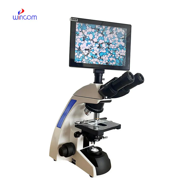
I’ve used several microscopes before, but this one stands out for its sturdy design and smooth magnification control.
This ultrasound scanner has truly improved our workflow. The image resolution and portability make it a great addition to our clinic.
To protect the privacy of our buyers, only public service email domains like Gmail, Yahoo, and MSN will be displayed. Additionally, only a limited portion of the inquiry content will be shown.
We’re interested in your delivery bed for our maternity department. Please send detailed specifica...
I’d like to inquire about your x-ray machine models. Could you provide the technical datasheet, wa...
E-mail: [email protected]
Tel: +86-731-84176622
+86-731-84136655
Address: Rm.1507,Xinsancheng Plaza. No.58, Renmin Road(E),Changsha,Hunan,China

 af
af
 es
es
 ar
ar
 tr
tr
 sw
sw
 pt
pt
 th
th
 ur
ur
 bn
bn
 ne
ne
 vi
vi
 km
km
 lo
lo
 de
de
 ru
ru
 fi
fi
 nl
nl
 fa
fa
 fr
fr
 ko
ko