The baby fetal doppler has been designed considering the needs of modern health care, providing uninterrupted performance with its rapid image acquisition and high-definition visualization. The robust outer casing and sophisticated temperature control system will make sure that the device continues to work and be trustworthy. Also, the baby fetal doppler is a device that aids the long-term data archiving process for efficient medical record management.

In emergency departments, the baby fetal doppler is used for instant imaging to easily spot internal wounds and bleeding. It supports the doctor with the abdominal trauma and chest condition diagnosis. Moreover, the baby fetal doppler provides assistance in rural and field medical practice, delivering consistent imaging in areas with poor medical facilities.

In addition, as new technologies emerge, the baby fetal doppler is expected to become more compact and intelligent with better diagnostic capabilities. The new baby fetal doppler will incorporate 3D and 4D capabilities. The baby fetal doppler will also be integrated with digital hospitals for seamless management of patient data.

Proper care and management of the baby fetal doppler needs to be carried out to ensure that it functions well at all times. Cleaning of the probes using a recommended disinfectant helps prevent contamination of the probe and image distortion. Storage of the baby fetal doppler in a clean and dry place away from high temperatures helps prolong the life of the equipment.
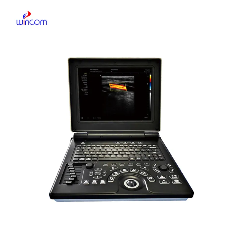
With the advanced imaging technology, the baby fetal doppler provides physicians unobstructed and precise images of internal organs. It is employed in the early disease diagnosis as well as in patient tracking. The baby fetal doppler functions by utilizing sound wave reflections to generate dynamic images, qualifying it as an essential tool in modern medical diagnostics. Through the baby fetal doppler, fast, non-invasive testing is facilitated for real-time assessment to support clinical decisions.
Q: What imaging modes are available on the ultrasound scannert? A: It supports multiple modes such as B-mode, M-mode, and color Doppler for diverse diagnostic applications. Q: How does the ultrasound scannert improve diagnostic accuracy? A: By providing high-resolution images and real-time feedback, it enables more precise medical evaluations. Q: Can the ultrasound scannert be used in field or remote settings? A: Yes, its portable versions are designed for mobility and can be used in clinics, hospitals, or mobile healthcare units. Q: What kind of display does the ultrasound scannert use? A: It typically features a high-definition digital display that enhances image visualization and readability. Q: How is data from the ultrasound scannert managed? A: The device allows secure storage, easy access, and export of imaging data through USB or network connections.
We’ve been using this mri machine for several months, and the image clarity is excellent. It’s reliable and easy for our team to operate.
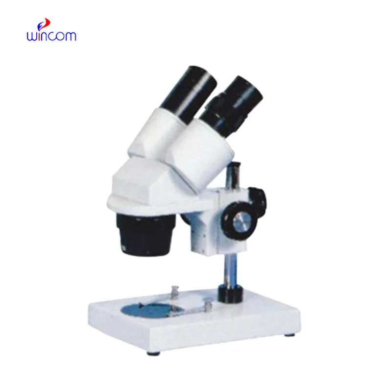
I’ve used several microscopes before, but this one stands out for its sturdy design and smooth magnification control.
To protect the privacy of our buyers, only public service email domains like Gmail, Yahoo, and MSN will be displayed. Additionally, only a limited portion of the inquiry content will be shown.
We are planning to upgrade our imaging department and would like more information on your mri machin...
Could you please provide more information about your microscope range? I’d like to know the magnif...
E-mail: [email protected]
Tel: +86-731-84176622
+86-731-84136655
Address: Rm.1507,Xinsancheng Plaza. No.58, Renmin Road(E),Changsha,Hunan,China

 af
af
 es
es
 ar
ar
 tr
tr
 sw
sw
 pt
pt
 th
th
 ur
ur
 bn
bn
 ne
ne
 vi
vi
 km
km
 lo
lo
 de
de
 ru
ru
 fi
fi
 nl
nl
 fa
fa
 fr
fr
 ko
ko