When it comes to durability and precision, the detector fetal doppler is a tough and highly accurate device that relies on the integration of the latest imaging software to provide quality diagnostic images. Its light structure and an electric system powered by rechargeable batteries make it a device that can be used at a stationary place or be taken out for mobile operations. The detector fetal doppler is a product that brings about a better workflow due to its easy-to-use controls and proper data management.

The detector fetal doppler is recognized for its great contribution to the field of surgery and thus is employed frequently in operating theaters for providing intraoperative guidance and ascertaining anatomical targets. It can easily locate areas where fluid has collected, determine the condition of the tissue, and provide evidence that the procedure has been successful. The detector fetal doppler can also be used dynamically and thus in sports medicine for imaging of muscles and tendons during movement analysis.

Through continued innovations in digital technology, the detector fetal doppler can be expected to improve and extend its applications within preventive medicine and telemedicine. The next generation of such technologies will facilitate collaboration among experts in real-time using cloud-imaging solutions. The detector fetal doppler can also work within wearables that include biosensors.

In order to extend the service life of the detector fetal doppler, it is recommended that users refrain from applying much force during the process of connecting/disconnecting probes. Power cables should always remain dry. The detector fetal doppler needs diagnostic tests to ensure that it produces quality images.
The detector fetal doppler uses state-of-the-art ultrasound technology to deliver real-time imaging for diagnostic and monitoring purposes. It aids physicians in assessing organs, blood vessels, and soft tissue with unmatched clarity. The non-surgical device is an important tool for guiding medical procedures and making precise diagnoses. The detector fetal doppler combines portability with precision, rendering it extremely useful in routine exams as well as emergency applications.
Q: What is the primary function of an ultrasound scannert? A: Ultrasound scanners are designed to create real-time images of internal organs, tissues, and blood flow using high-frequency sound waves. Q: How does the ultrasound scannert ensure clear imaging results? A:It uses advanced converter technology and digital processing to enhance image clarity and contrast. Q: In what medical fields is the ultrasound scannert commonly used? A: It is widely used in obstetrics, cardiology, urology, radiology, and emergency medicine. Q: Is the ultrasound scannert safe for repeated use? A: Yes, it is non-invasive and does not emit radiation, making it safe for frequent diagnostic applications. Q: Can the ultrasound scannert store and share imaging data? A: Yes, it supports data storage, retrieval, and digital transfer for easy integration with hospital systems.
This ultrasound scanner has truly improved our workflow. The image resolution and portability make it a great addition to our clinic.
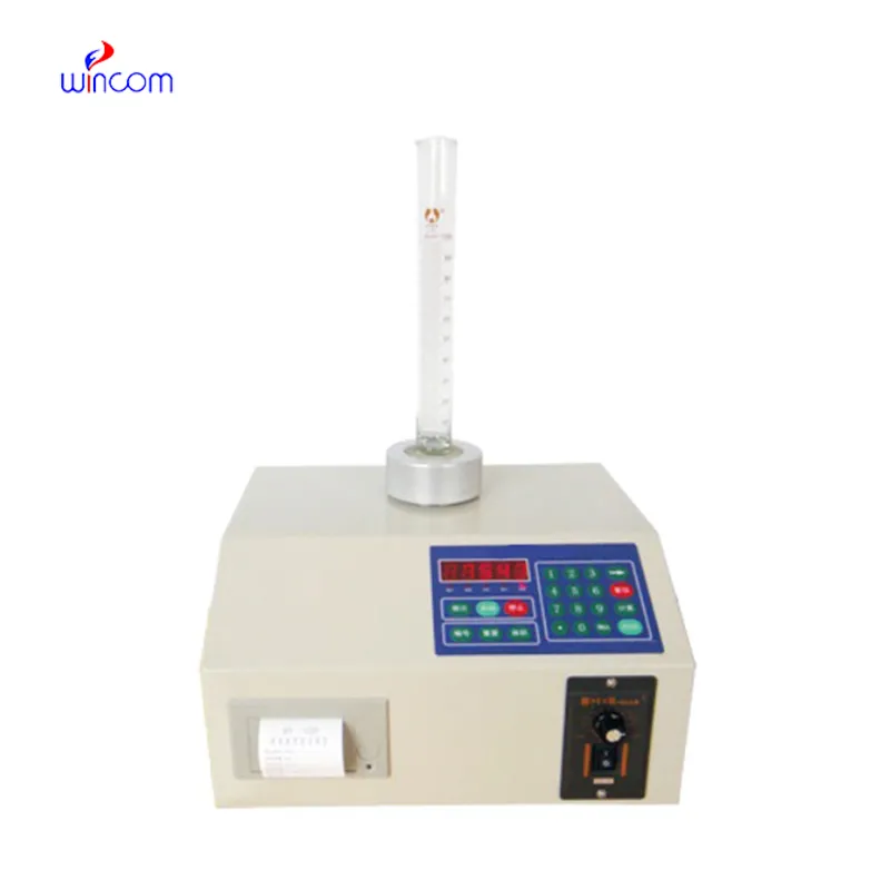
The centrifuge operates quietly and efficiently. It’s compact but surprisingly powerful, making it perfect for daily lab use.
To protect the privacy of our buyers, only public service email domains like Gmail, Yahoo, and MSN will be displayed. Additionally, only a limited portion of the inquiry content will be shown.
I’d like to inquire about your x-ray machine models. Could you provide the technical datasheet, wa...
We’re interested in your delivery bed for our maternity department. Please send detailed specifica...
E-mail: [email protected]
Tel: +86-731-84176622
+86-731-84136655
Address: Rm.1507,Xinsancheng Plaza. No.58, Renmin Road(E),Changsha,Hunan,China

 af
af
 es
es
 ar
ar
 tr
tr
 sw
sw
 pt
pt
 th
th
 ur
ur
 bn
bn
 ne
ne
 vi
vi
 km
km
 lo
lo
 de
de
 ru
ru
 fi
fi
 nl
nl
 fa
fa
 fr
fr
 ko
ko