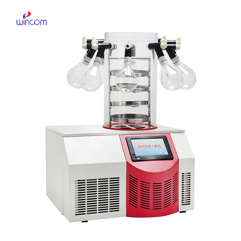
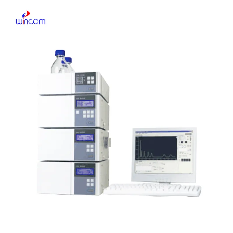
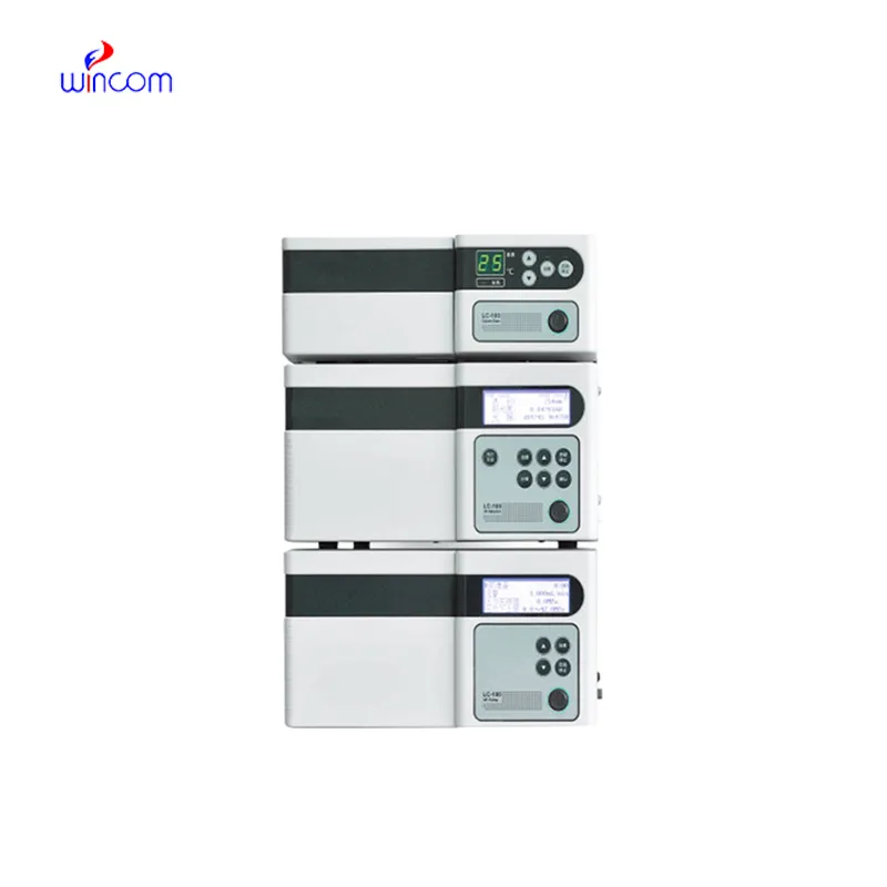
Today, clinical laboratories always rely on guard column hplc for the purpose of giving comprehensive chemical and biological data from patient samples. The technology's exceptional sensitivity and accuracy make it possible to separate even the smallest amounts of substances such as drugs and metabolites from complicated mixtures. Laboratory staff performs using guard column hplc in method development, validation and ongoing monitoring of the lab's analytical performance. The multi-use of the instrument guarantees its presence during both normal testing and research work, hence hospitals and laboratories are always consistent in providing accurate and trustworthy diagnostic and analytical results.

The quality control process for guard column hplc in intravenous medications and hospital-prepared solutions is being carried out by hospital laboratories. It isolates the impurities and analyzes the active substances to ascertain the uniformity of the composition. This practice enables the pharmacists and laboratory staff to verify the drug's quality before it gets to the patient, hence minimizing the risk associated with it and at the same time endorsing the safe therapeutic practices in hospitals.
In hospitals and clinical research, guard column hplc techniques will get higher resolution columns and ultrafast chromatography methods more and more. It will be possible to do these innovations in a shorter time and with a more accurate result. Future guard column hplc applications will be used to identify biomarkers quickly, monitor therapies in real-time, and manage patients more efficiently in both the laboratory and clinical settings.
Routine upkeep of guard column hplc is of utmost importance in clinical laboratories to maintain the accuracy of patient sample analysis. Regular cleaning of pipes, changing of deteriorated seals and calibration of measuring instruments will block adulteration and keep the latter's sensitivity. Lab personnel must record maintenance activities and keep watch over system performance. Constant attention guarantees that guard column hplc provides dependable, reproducible results for hospital diagnosis and research work.
Clinical laboratories make use of guard column hplc to analyze patient samples with remarkable accuracy. It identifies biomarkers, metabolites, and the levels of therapeutic drugs, thus giving reliable information about the disease status and monitoring treatment. Sensitivity of the technique permits determination of compounds in very minute amounts, which is critical in clinical testing. By resolving complex composition, guard column hplc guarantees accurate and reproducible results for laboratory diagnostics. Lab staff utilizes it for daily testing, quality control, and research activities, thus making guard column hplc a vital part of contemporary clinical laboratory work that caters to patient care, treatment choices, and lab data integrity.
Q: Do you need special training for HPLC operation? A: The answer is yes, training is a prerequisite to accurately and safely using pumps, columns, and detectors. Q: What type of maintenance does HPLC have? A: It requires cleaning, flushing, and inspection of all components as well as calibrating. Q: Is it possible to use HPLC in drug monitoring? A: Sure, it is a common practice in hospitals to monitor the levels of therapeutic drugs and also to identify metabolites in the samples taken from the patients. Q: What is the duration of analysis using HPLC in a typical case? A: The analysis time can range from a few minutes to more than an hour depending on the nature of the sample and the kind of column used. Q: Is HPLC a good choice for environmental testing? A: Yes, it can be used to find out the presence of pollutants, pesticides, and other harmful substances in water, soil, and air samples.
The centrifuge operates quietly and efficiently. It’s compact but surprisingly powerful, making it perfect for daily lab use.
The water bath performs consistently and maintains a stable temperature even during long experiments. It’s reliable and easy to operate.
To protect the privacy of our buyers, only public service email domains like Gmail, Yahoo, and MSN will be displayed. Additionally, only a limited portion of the inquiry content will be shown.
We’re currently sourcing an ultrasound scanner for hospital use. Please send product specification...
We’re looking for a reliable centrifuge for clinical testing. Can you share the technical specific...
E-mail: [email protected]
Tel: +86-731-84176622
+86-731-84136655
Address: Rm.1507,Xinsancheng Plaza. No.58, Renmin Road(E),Changsha,Hunan,China

 af
af
 es
es
 ar
ar
 tr
tr
 sw
sw
 pt
pt
 th
th
 ur
ur
 bn
bn
 ne
ne
 vi
vi
 km
km
 lo
lo
 de
de
 ru
ru
 fi
fi
 nl
nl
 fa
fa
 fr
fr
 ko
ko