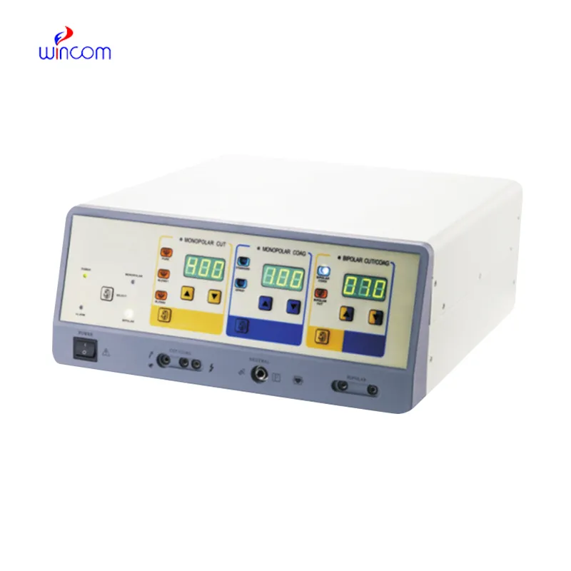
The home fetal heart doppler, being designed for flexibility, brings the user a variety of imaging modes such as B-mode, M-mode, and color Doppler to use. Because of its small size, it can be easily moved from one place to another and that is why it can be used for examining patients in bed. The home fetal heart doppler provides good imaging quality, which can be depended upon in cases of routine diagnostics, fieldwork, and emergency medical interventions.

The home fetal heart doppler is a very significant diagnosis tool used in obstetrics for fetal monitoring and pregnancy development detection. It indirectly affects the cardiology field by providing information about the hearts and blood flow dynamics. Furthermore, the home fetal heart doppler is very important in diagnosing abdominal problems, especially issues with the liver, kidneys, and gallbladder. Still, it is also being used in musculoskeletal diagnoses for spotting ligament and tendon injuries.

The home fetal heart doppler should integrate with intelligent diagnostic ecosystems and communicate effortlessly with smartphones and electronic records. The synchronized exchange of data in real-time should enable constant patient observation. The next version should focus on improved design, better processing power of artificial intelligence algorithms, and enhanced reconstruction functions.

In order to retain the accuracy of the home fetal heart doppler, it is important for operators to check the cables and connections of the transducers for evidence of wear. After each use, the surfaces should be wiped clean using non-abrasive cleaners. The home fetal heart doppler should be turned off properly and covered to prevent dust from collecting. Regular checks by trained personnel should be done.
Designed to be accurate and functional, the home fetal heart doppler offers high-definition imaging for diagnosis in numerous medical settings. It is comfortable with obstetric, vascular, and abdominal procedures and delivers exceptional definition. The home fetal heart doppler increases the certainty of diagnosis and reduces patient disruption through its non-invasive mode of operation. Its digital components allow for storage of data, transfer of images, and analysis.
Q: What imaging modes are available on the ultrasound scannert? A: It supports multiple modes such as B-mode, M-mode, and color Doppler for diverse diagnostic applications. Q: How does the ultrasound scannert improve diagnostic accuracy? A: By providing high-resolution images and real-time feedback, it enables more precise medical evaluations. Q: Can the ultrasound scannert be used in field or remote settings? A: Yes, its portable versions are designed for mobility and can be used in clinics, hospitals, or mobile healthcare units. Q: What kind of display does the ultrasound scannert use? A: It typically features a high-definition digital display that enhances image visualization and readability. Q: How is data from the ultrasound scannert managed? A: The device allows secure storage, easy access, and export of imaging data through USB or network connections.
We’ve used this centrifuge for several months now, and it has performed consistently well. The speed control and balance are excellent.
The delivery bed is well-designed and reliable. Our staff finds it simple to operate, and patients feel comfortable using it.
To protect the privacy of our buyers, only public service email domains like Gmail, Yahoo, and MSN will be displayed. Additionally, only a limited portion of the inquiry content will be shown.
I’d like to inquire about your x-ray machine models. Could you provide the technical datasheet, wa...
I’m looking to purchase several microscopes for a research lab. Please let me know the price list ...
E-mail: [email protected]
Tel: +86-731-84176622
+86-731-84136655
Address: Rm.1507,Xinsancheng Plaza. No.58, Renmin Road(E),Changsha,Hunan,China

 af
af
 es
es
 ar
ar
 tr
tr
 sw
sw
 pt
pt
 th
th
 ur
ur
 bn
bn
 ne
ne
 vi
vi
 km
km
 lo
lo
 de
de
 ru
ru
 fi
fi
 nl
nl
 fa
fa
 fr
fr
 ko
ko