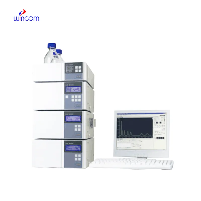
hplc analyse is a critical technique to obtain analytical information in studies of medicines, clinical samples, and biochemistry. It isolates compounds according to their chemical characteristics, generating reproducible analytical results. Laboratory scientists use hplc analyse to perform drug stability tests, monitor patient biomarkers, and find impurities. Its very high accuracy and flexibility allow thorough sample analysis in research, hospital, and clinical laboratory environments, thus becoming a fundamental device for assuring precision in both experimental and diagnostic results.

hplc analyse are utilized by clinical laboratories for hormone and endocrine-related biomarker detection. It delivers trustworthy information for the diagnosis of endocrine diseases by correctly separating substances like cortisol, thyroid hormones, or insulin. Techs in a laboratory rely on hplc analyse to give accurate and repeatable results, thereby helping doctors in individual treatment plan.
Hospital laboratories will largely benefit from hplc analyse systems that are meant for increased throughput and multi-sample analysis. The future instruments will merge improved sensitivity with strong automation, thus making rapid diagnostics and continuous monitoring of patient medications and metabolic profiles possible, which in turn will provide hospitals with safer and more efficient operations.
hplc analyse will require regular maintenance to be kept up in order to continue providing precise measurements in medical laboratories. After every use, the technicians should flush the columns, check the seals, and inspect the tubing for wear and tear and ensure that the detector is working. Regular calibration and good solvent management decrease the chances of system damage and increase the consistency of the results. Good care and maintenance not only increase the efficiency of the laboratory but also help in providing reliable diagnostics and maintaining the instruments for hospital applications.
hplc analyse is of utmost importance in biochemistry laboratories of both universities and hospitals. It makes detailed study of proteins, peptides, and metabolites possible through the separation of intricate mixtures. The application of it includes but is not limited to enzymatic analysis, biomarker detection, and data obtained through metabolomics. The sensitivity and reproducibility of the device guarantee genuine molecular profiles. Lab technicians make use of hplc analyse to conclude their experiments and provide evidence for scientific publications. Its accuracy and versatility give biochemistry labs the ability to perform cutting-edge research in molecular mechanisms, disease pathways, and therapy targets thus, it becomes an indispensable tool for both analytical and clinical lab investigations.
Q: What is the sample preparation for HPLC? A: For the most part, samples should be filtered, diluted, or subjected to solvent extraction in order to avoid column clogs and have the results be accurate Q: Is HPLC able to pick trace-level compounds? A: With the right detectors, it can pick up such substances in extremely small amounts with high sensitivity. Q: Is HPLC a method that can be applied to analysis of proteins? A: Yes, particularly if one employs size-exclusion and reversed-phase columns for protein, peptide, and biomolecule separation. Q: What is the process of calibrating HPLC? A: The process is done by taking standards of known concentrations that are the same as the one in the sample and using them to check the performance of the column and the accuracy of the detector. Q: Are particular solvents needed for HPLC? A: Yes, the solvents used need to be compatible with the type of the column and the detectors to prevent any damage or interference in the analysis process.
We’ve been using this mri machine for several months, and the image clarity is excellent. It’s reliable and easy for our team to operate.
The centrifuge operates quietly and efficiently. It’s compact but surprisingly powerful, making it perfect for daily lab use.
To protect the privacy of our buyers, only public service email domains like Gmail, Yahoo, and MSN will be displayed. Additionally, only a limited portion of the inquiry content will be shown.
I’d like to inquire about your x-ray machine models. Could you provide the technical datasheet, wa...
Could you please provide more information about your microscope range? I’d like to know the magnif...
E-mail: [email protected]
Tel: +86-731-84176622
+86-731-84136655
Address: Rm.1507,Xinsancheng Plaza. No.58, Renmin Road(E),Changsha,Hunan,China

 af
af
 es
es
 ar
ar
 tr
tr
 sw
sw
 pt
pt
 th
th
 ur
ur
 bn
bn
 ne
ne
 vi
vi
 km
km
 lo
lo
 de
de
 ru
ru
 fi
fi
 nl
nl
 fa
fa
 fr
fr
 ko
ko