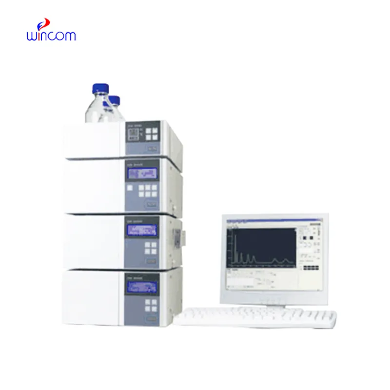
hplc def is a critical technique to obtain analytical information in studies of medicines, clinical samples, and biochemistry. It isolates compounds according to their chemical characteristics, generating reproducible analytical results. Laboratory scientists use hplc def to perform drug stability tests, monitor patient biomarkers, and find impurities. Its very high accuracy and flexibility allow thorough sample analysis in research, hospital, and clinical laboratory environments, thus becoming a fundamental device for assuring precision in both experimental and diagnostic results.

Hospital laboratories depend on hplc def for identifying minute quantities of pharmaceuticals and therapeutic agents in difficult-to-analyze biological samples. Its use spans drug compliance testing, pharmacokinetics profiling, and tracking medications after surgery. The laboratory personnel can rely on it for exact measurement, thus increasing the efficiency of clinical treatment.
hplc def is assigned to become an important player in translational research which is being conducted in hospitals. Among the future developments are the combined detection systems, quicker analysis cycles, and improved reproducibility. hplc def will be the mainstay of hospitals' molecular profiling and drug testing along with patient monitoring thus facilitating hospital diagnostics and personalized medicine research.
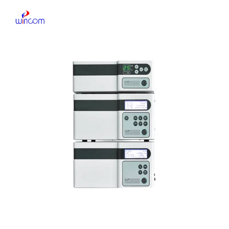
Regular system checks, cleaning of detector flow cells, and changing consumable parts whenever necessary are some of the actions that the laboratory staff should take in order to keep the hplc def working efficiently. Observing pump performance, taking care of solvent contamination, and storing columns correctly prolong the life of the instrument. Good maintenance assures reproducibility, cuts down on time without access to equipment, and promotes high-quality analysis in hospitals and clinical labs.
Therapeutic drug monitoring relies heavily on hplc def in hospital settings. It determines the concentration of drugs in the body to guarantee efficiency and security. The laboratory staff uses it for the examination of blood, serum, or urine samples, and signifies small molecular compounds with high accuracy. By yielding consistent outcomes, hplc def services the medics in changing the amounts and preventing side effects. Its use goes to hormone level testing, metabolite analysis, and pharmacokinetics research. With quick processing and accurate information, hplc def is a part of the hospital patient care, making evidence-based treatment decisions possible and enhancing clinical outcomes in different departments.
Q: What are the main parts of a microscope? A: The key components include the eyepiece, objective lenses, stage, focusing knobs, and illumination system, all working together to magnify and clarify specimens. Q: How do you clean the lenses of a microscope? A: Lenses should be cleaned using soft lens paper or microfiber cloth with a small amount of lens cleaner to avoid scratching or damaging optical coatings. Q: What magnification levels can a microscope achieve? A: Depending on the model, a microscope can typically achieve magnifications ranging from 40x to over 1000x for detailed observation of microscopic structures. Q: Why is light adjustment important in a microscope? A: Proper light adjustment ensures accurate contrast and brightness, allowing clear observation without distortion or glare during viewing. Q: Can a microscope be used for educational purposes? A: Yes, microscopes are widely used in classrooms and laboratories to teach students about biology, materials science, and microscopic analysis.
The microscope delivers incredibly sharp images and precise focusing. It’s perfect for both professional lab work and educational use.
The hospital bed is well-designed and very practical. Patients find it comfortable, and nurses appreciate how simple it is to operate.
To protect the privacy of our buyers, only public service email domains like Gmail, Yahoo, and MSN will be displayed. Additionally, only a limited portion of the inquiry content will be shown.
Could you please provide more information about your microscope range? I’d like to know the magnif...
I’d like to inquire about your x-ray machine models. Could you provide the technical datasheet, wa...
E-mail: [email protected]
Tel: +86-731-84176622
+86-731-84136655
Address: Rm.1507,Xinsancheng Plaza. No.58, Renmin Road(E),Changsha,Hunan,China

 af
af
 es
es
 ar
ar
 tr
tr
 sw
sw
 pt
pt
 th
th
 ur
ur
 bn
bn
 ne
ne
 vi
vi
 km
km
 lo
lo
 de
de
 ru
ru
 fi
fi
 nl
nl
 fa
fa
 fr
fr
 ko
ko