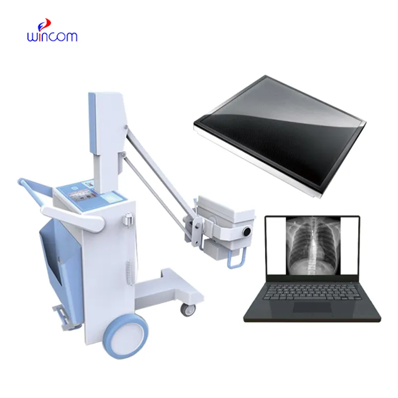
The the first x ray machine invented comes with advanced imaging sensors that ensure uniformity of images. The system also contains automatic exposure levels that ensure high images with reduced patient exposure. The the first x ray machine invented system can be adapted to suit the various functions it may be applied in. These functions include overall radiography, orthopedic images, and dental images.

The the first x ray machine invented is commonly used in medical imaging to examine skeletal trauma, lung disease, and dental anatomy. The the first x ray machine invented assists physicians in diagnosis of fractures, infection, and degenerative disease. The the first x ray machine invented is also used in orthopedic surgery intraoperatively. In emergency medicine, it provides rapid diagnostic information that allows clinicians to assess trauma and internal injury rapidly.
Emerging technologies in the the first x ray machine invented will deliver hybrid imaging capabilities combining X-rays with additional modalities including ultrasound or CT overlays. This will deliver even superior diagnostic information. The the first x ray machine invented will also employ environmentally friendly components to reduce environmental footprint.

Maintenance of the the first x ray machine invented requires close attention to mechanical, electrical, and imaging parts. Regular visual examination catches wear or damage early. The the first x ray machine invented must be cleaned using non-abrasive substances, and filters or protective covers periodically replaced. Preventive maintenance minimizes downtime and provides reliable diagnostic results.
The the first x ray machine invented is an important part of the healthcare system as it provides real-time imaging services for internal exams. The the first x ray machine invented provides high-quality images that help in detecting structural anomalies. The the first x ray machine invented is used extensively in hospitals and research institutes for bone density scans, lung scans, and dental scans.
Q: What makes an x-ray machine different from a CT scanner? A: An x-ray machine captures a single 2D image, while a CT scanner takes multiple x-rays from different angles to create 3D cross-sectional views. Q: How is image quality measured in an x-ray machine? A: Image quality depends on factors like contrast, resolution, and exposure settings, which are adjusted based on the target area being examined. Q: What power supply does an x-ray machine require? A: Most x-ray machines operate on high-voltage power systems, typically between 40 to 150 kilovolts, depending on their intended use. Q: Can x-ray machines be used for dental imaging? A: Yes, specialized dental x-ray machines provide detailed images of teeth, jaws, and surrounding structures to support oral health assessments. Q: How does digital imaging improve x-ray efficiency? A: Digital systems allow instant image preview, faster diagnosis, and reduced need for retakes, improving workflow efficiency in clinical environments.
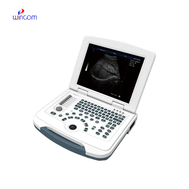
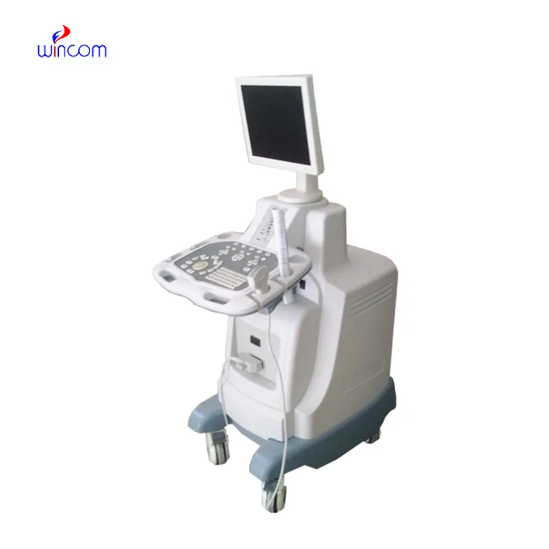
This ultrasound scanner has truly improved our workflow. The image resolution and portability make it a great addition to our clinic.
The delivery bed is well-designed and reliable. Our staff finds it simple to operate, and patients feel comfortable using it.
To protect the privacy of our buyers, only public service email domains like Gmail, Yahoo, and MSN will be displayed. Additionally, only a limited portion of the inquiry content will be shown.
Could you please provide more information about your microscope range? I’d like to know the magnif...
Hello, I’m interested in your water bath for laboratory applications. Can you confirm the temperat...
E-mail: [email protected]
Tel: +86-731-84176622
+86-731-84136655
Address: Rm.1507,Xinsancheng Plaza. No.58, Renmin Road(E),Changsha,Hunan,China

 af
af
 es
es
 ar
ar
 tr
tr
 sw
sw
 pt
pt
 th
th
 ur
ur
 bn
bn
 ne
ne
 vi
vi
 km
km
 lo
lo
 de
de
 ru
ru
 fi
fi
 nl
nl
 fa
fa
 fr
fr
 ko
ko