The ultrasound 3d scanner has been designed considering the needs of modern health care, providing uninterrupted performance with its rapid image acquisition and high-definition visualization. The robust outer casing and sophisticated temperature control system will make sure that the device continues to work and be trustworthy. Also, the ultrasound 3d scanner is a device that aids the long-term data archiving process for efficient medical record management.

The ultrasound 3d scanner is recognized for its great contribution to the field of surgery and thus is employed frequently in operating theaters for providing intraoperative guidance and ascertaining anatomical targets. It can easily locate areas where fluid has collected, determine the condition of the tissue, and provide evidence that the procedure has been successful. The ultrasound 3d scanner can also be used dynamically and thus in sports medicine for imaging of muscles and tendons during movement analysis.

The ultrasound 3d scanner will proceed to develop as new innovations emerge in artificial intelligence and data analysis. The new models of the ultrasound 3d scanner will be able to provide training simulations that experts can use to improve scanning sessions. The increased processing power and connectivity of the ultrasound 3d scanner will set new standards of accessibility and accuracy in medical scanning.

For long-term functionality, it is recommended that the ultrasound 3d scanner remain within an environment that maintains controlled levels of both humidity and temperature. The cables should be unwound slowly to ensure that no undue stress or wire breakages occur. The ultrasound 3d scanner should also be properly disinfected each time a patient has been examined.
The ultrasound 3d scanner is a critical diagnostic tool used across all the medical specialties for imaging internal organs, tissues, and blood flow in real time. It generates high-resolution images with no patient radiation exposure using high-frequency sound waves. The ultrasound 3d scanner ensures precise monitoring across obstetrics, cardiology, and emergency medicine. Its portability and simplicity ensure it is useful in both field and clinical settings, enhancing diagnostic efficiency and precision.
Q: What is the primary function of an ultrasound scannert? A: Ultrasound scanners are designed to create real-time images of internal organs, tissues, and blood flow using high-frequency sound waves. Q: How does the ultrasound scannert ensure clear imaging results? A:It uses advanced converter technology and digital processing to enhance image clarity and contrast. Q: In what medical fields is the ultrasound scannert commonly used? A: It is widely used in obstetrics, cardiology, urology, radiology, and emergency medicine. Q: Is the ultrasound scannert safe for repeated use? A: Yes, it is non-invasive and does not emit radiation, making it safe for frequent diagnostic applications. Q: Can the ultrasound scannert store and share imaging data? A: Yes, it supports data storage, retrieval, and digital transfer for easy integration with hospital systems.
The water bath performs consistently and maintains a stable temperature even during long experiments. It’s reliable and easy to operate.
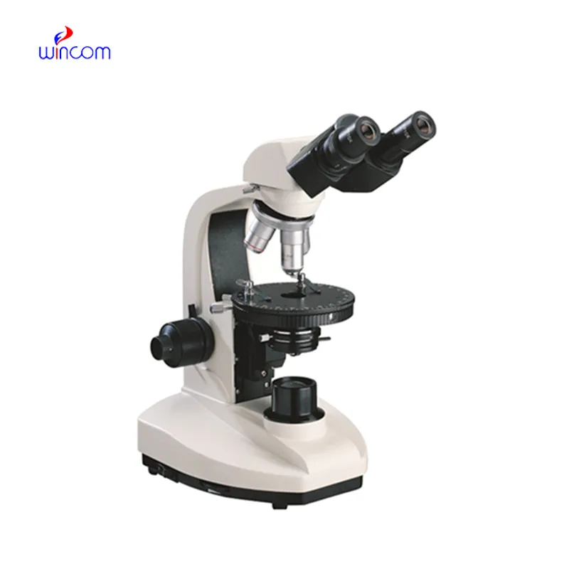
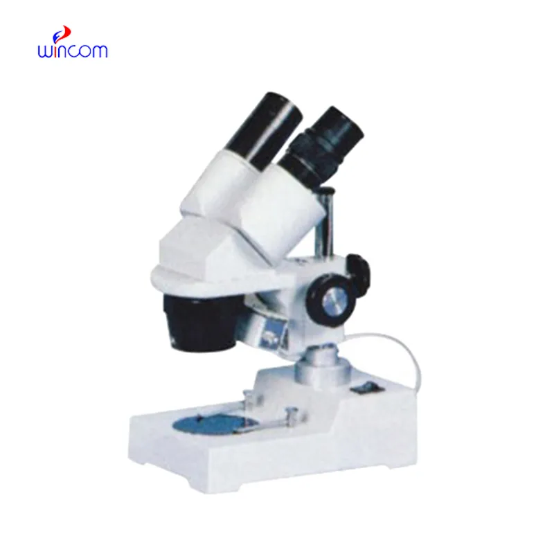
I’ve used several microscopes before, but this one stands out for its sturdy design and smooth magnification control.
To protect the privacy of our buyers, only public service email domains like Gmail, Yahoo, and MSN will be displayed. Additionally, only a limited portion of the inquiry content will be shown.
I’d like to inquire about your x-ray machine models. Could you provide the technical datasheet, wa...
Hello, I’m interested in your water bath for laboratory applications. Can you confirm the temperat...
E-mail: [email protected]
Tel: +86-731-84176622
+86-731-84136655
Address: Rm.1507,Xinsancheng Plaza. No.58, Renmin Road(E),Changsha,Hunan,China

 af
af
 es
es
 ar
ar
 tr
tr
 sw
sw
 pt
pt
 th
th
 ur
ur
 bn
bn
 ne
ne
 vi
vi
 km
km
 lo
lo
 de
de
 ru
ru
 fi
fi
 nl
nl
 fa
fa
 fr
fr
 ko
ko