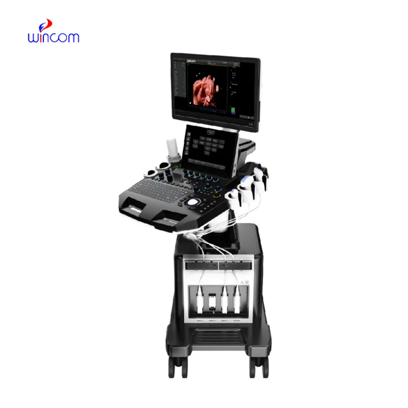
Using state-of-the-art real-time signal processing, the ultrasound scanner has the capability of producing imaging output that is invariably sharp. The system of the device is capable of dynamically tuning the frequency and gain for achieving the best image quality. The ultrasound scanner with its versatile probe compatibility is able to deal with different demanding clinical applications like obstetrics, cardiology, and abdominal scans.

The ultrasound scanner has a very wide use in radiology where it supports the guidance of non-invasive procedures. It is very important in gynecology where it is allowed to conduct reproductive system evaluations. In orthopedics, the ultrasound scanner helps visualize muscles, joints, and tendons ensuring correct diagnostic interpretation. It is this very nature of versatility that makes it a must-have for imaging during procedures in real time.

In addition, as new technologies emerge, the ultrasound scanner is expected to become more compact and intelligent with better diagnostic capabilities. The new ultrasound scanner will incorporate 3D and 4D capabilities. The ultrasound scanner will also be integrated with digital hospitals for seamless management of patient data.

The daily upkeep of the ultrasound scanner involves cleaning, inspection, and proper storage. The removal of the gel residue from the probes should be accomplished as soon as the analysis has been carried out. The cooling vents of the device should always be unblocked. The ultrasound scanner needs annual professional servicing in order to remain accurate.
The ultrasound scanner is more accurate in diagnostics as it captures high-resolution images of organs, tissues, and blood vessels. Design-wise flexible, it is used extensively in obstetrics, cardiology, urology, and musculoskeletal tests. Its portability and simplicity enable medical practitioners to make quick and precise evaluations. The ultrasound scanner makes work processes more efficient and allows for the delivery of superior patient care through real-time visualization.
Q: What imaging modes are available on the ultrasound scannert? A: It supports multiple modes such as B-mode, M-mode, and color Doppler for diverse diagnostic applications. Q: How does the ultrasound scannert improve diagnostic accuracy? A: By providing high-resolution images and real-time feedback, it enables more precise medical evaluations. Q: Can the ultrasound scannert be used in field or remote settings? A: Yes, its portable versions are designed for mobility and can be used in clinics, hospitals, or mobile healthcare units. Q: What kind of display does the ultrasound scannert use? A: It typically features a high-definition digital display that enhances image visualization and readability. Q: How is data from the ultrasound scannert managed? A: The device allows secure storage, easy access, and export of imaging data through USB or network connections.
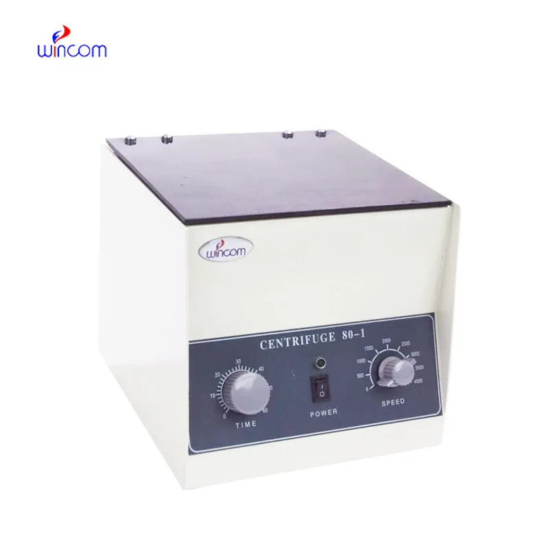
The centrifuge operates quietly and efficiently. It’s compact but surprisingly powerful, making it perfect for daily lab use.
We’ve been using this mri machine for several months, and the image clarity is excellent. It’s reliable and easy for our team to operate.
To protect the privacy of our buyers, only public service email domains like Gmail, Yahoo, and MSN will be displayed. Additionally, only a limited portion of the inquiry content will be shown.
We are planning to upgrade our imaging department and would like more information on your mri machin...
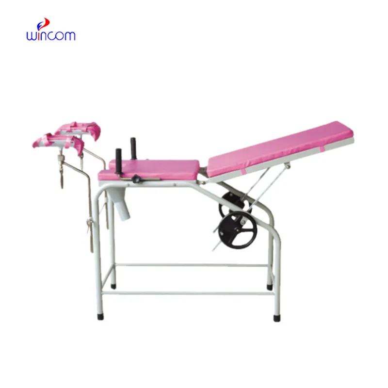
We’re interested in your delivery bed for our maternity department. Please send detailed specifica...
E-mail: [email protected]
Tel: +86-731-84176622
+86-731-84136655
Address: Rm.1507,Xinsancheng Plaza. No.58, Renmin Road(E),Changsha,Hunan,China

 af
af
 es
es
 ar
ar
 tr
tr
 sw
sw
 pt
pt
 th
th
 ur
ur
 bn
bn
 ne
ne
 vi
vi
 km
km
 lo
lo
 de
de
 ru
ru
 fi
fi
 nl
nl
 fa
fa
 fr
fr
 ko
ko