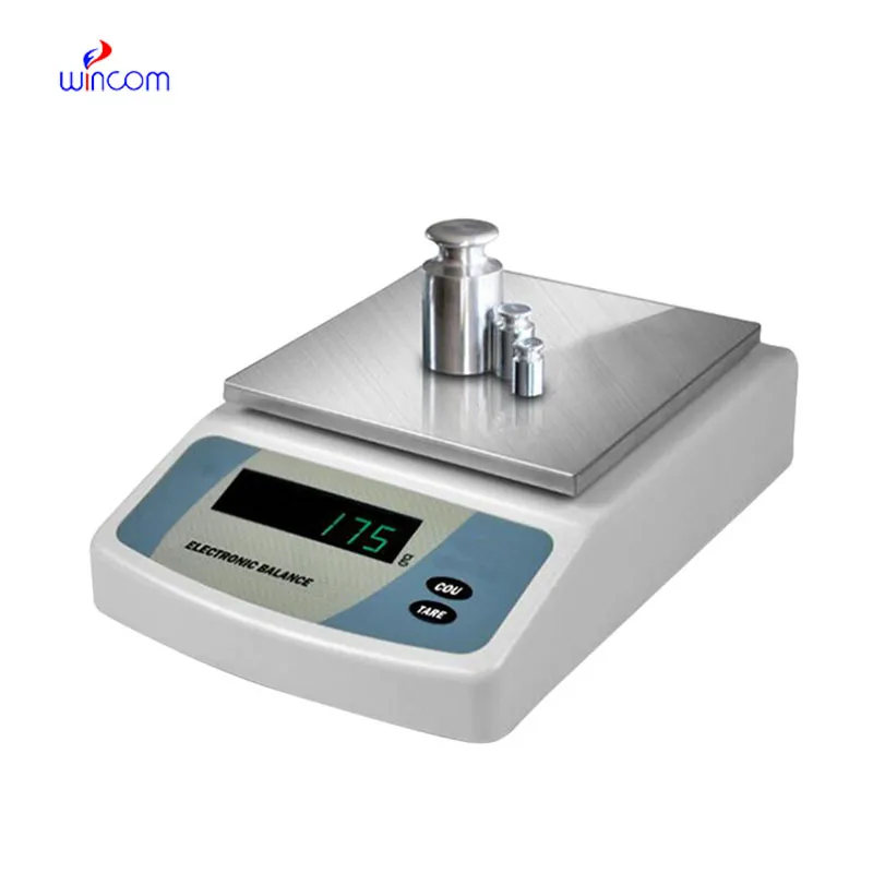
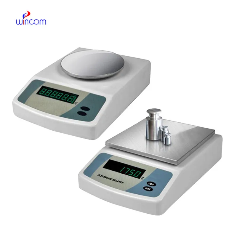
In the laboratories of hospitals and clinics, using an electronic balance is responsible for the precise weighing of patient samples, reagents, and pharmaceutical ingredients. Being highly precise, it minimized sample preparation errors and that is a good support for analytical results that can be reproduced. Laboratory techs apply using an electronic balance in the processes of quality control, method validation, and even daily operations. Reliable diagnostics, efficient laboratory workflows, and high-quality research and medical testing are the consequences of the accuracy and consistency maintained by using an electronic balance.

In pathology laboratories, using an electronic balance finds its usage during the staining compounds preparation and the tissue processing additives application. The proper mass measurement guarantees the same composition of the reagent, and this, in turn, affects the performance of the stain and the interpretation under the microscope. This application helps to maintain the same standards in pathology workflows and minimizes the differences between test batches. By using strict preparation conditions, using an electronic balance plays a role in the stability of diagnosis in hospital pathology departments.
In forthcoming times, using an electronic balance is foreseen to assimilate additional digital interrelation among hospital laboratories. Improved information interchange will make it possible to transfer weighing records directly to laboratory information systems, thus cutting down paper work done manually. This advancement will contribute to traceability, audit readiness, and efficiency in clinical workflows. When hospitals keep on embracing intelligent laboratory infrastructure, using an electronic balance will be an essential element of connected analytical ecosystems, helping with real-time monitoring and centralized data management.
using an electronic balance in clinics is, however, maintained through regular performance verification conducted in the laboratories. Certified test weights are used to prove the reliability of the measurements with the passage of time. Verification activities being documented also implies good traceability and makes it easier for internal audits to take place. By making verification part of the routine maintenance, hospitals make sure that using an electronic balance still gives trustworthy results for research and diagnostic workflows.
using an electronic balance plays an important role in the hospital pharmacy in the accurate formulation of medications, intravenous solutions, and compounded drugs. Even slight alterations in the weight of drugs can change the effectiveness of the drug and endanger the patient's safety. The Pharmacy Technicians rely on using an electronic balance for the correct dosing and checking of the active ingredients. The tool's accuracy guarantees the dependable preparation of drugs, the observance of the rules, and the quality control of the overall hospital pharmacy activities.
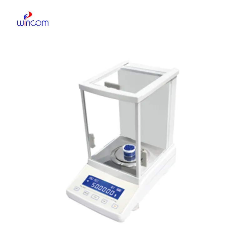
Q: What distinguishes an Analytical Balance from a precision balance? A: The analytical balances have a higher sensitivity and a finer readability for measuring masses of very small amounts. Q: Is an Analytical Balance appropriate for pharmaceutical applications? A: It is widely used for weighing active ingredient and formulation components. Q: Is it mandatory for an Analytical Balance to have a draft shield? A: Draft shields have the function to prevent air disturbances which might affect the weighing results. Q: What are the possible types of materials that can be weighed on an Analytical Balance? A: Weighing of powders, chemicals, and biological samples, as well as reference weights are the most common measurement. Q: Is it possible for several users to work with the same Analytical Balance? A: Yes, but the proper handling procedures and access controls must be strictly adhered to.
This ultrasound scanner has truly improved our workflow. The image resolution and portability make it a great addition to our clinic.
The centrifuge operates quietly and efficiently. It’s compact but surprisingly powerful, making it perfect for daily lab use.
To protect the privacy of our buyers, only public service email domains like Gmail, Yahoo, and MSN will be displayed. Additionally, only a limited portion of the inquiry content will be shown.
I’m looking to purchase several microscopes for a research lab. Please let me know the price list ...
Hello, I’m interested in your water bath for laboratory applications. Can you confirm the temperat...
E-mail: [email protected]
Tel: +86-731-84176622
+86-731-84136655
Address: Rm.1507,Xinsancheng Plaza. No.58, Renmin Road(E),Changsha,Hunan,China

 af
af
 es
es
 ar
ar
 tr
tr
 sw
sw
 pt
pt
 th
th
 ur
ur
 bn
bn
 ne
ne
 vi
vi
 km
km
 lo
lo
 de
de
 ru
ru
 fi
fi
 nl
nl
 fa
fa
 fr
fr
 ko
ko