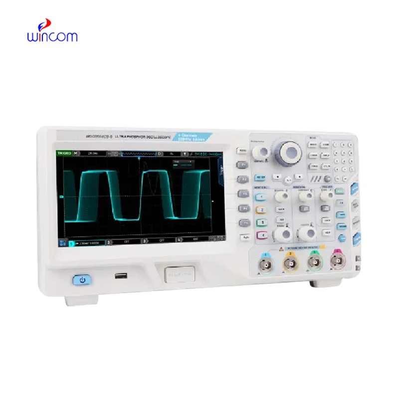
The x ray machine images comes equipped with advanced digital detectors that transform X-ray energy into high definition images of incredible detail. The system's design makes it easy to use and facilitates quick image capturing. The x ray machine images system can be connected effortlessly to hospital information systems that enable the secure transfer of data. The system's robust design provides support for long-term use within healthcare settings.

The x ray machine images is commonly used in medical imaging to examine skeletal trauma, lung disease, and dental anatomy. The x ray machine images assists physicians in diagnosis of fractures, infection, and degenerative disease. The x ray machine images is also used in orthopedic surgery intraoperatively. In emergency medicine, it provides rapid diagnostic information that allows clinicians to assess trauma and internal injury rapidly.

The x ray machine images will move further forward with advances in detector materials and digital processing. Future systems will provide better image quality at much lower radiation doses. With more advanced AI-assisted workflows, the x ray machine images will enable radiologists to spend more time on clinical interpretation and less on hand-tweaking.

To extend the life of the x ray machine images, it is recommended that operators follow maintenance procedures, including cleaning the X-ray tube housing and ensuring alignment accuracy. The x ray machine images may be turned off when not in use and protected from voltage fluctuations. Periodic quality assurance testing ensures image accuracy and patient safety.
Through the use of high-tech detectors and digital imaging, the x ray machine images provides high-quality internal structural images. The device enables healthcare providers to track various conditions such as pneumonia, arthritis, and dental cavities. The x ray machine images offers accurate imaging and ease of handling that makes it imperative in diagnostic radiology.
Q: What is an x-ray machine used for? A: An x-ray machine is used to produce images of the internal structures of the body, helping doctors detect fractures, infections, and other medical conditions. Q: How does an x-ray machine work? A:X-ray machine emit controlled radiation that passes through the body and records varying degrees of absorption on detectors or film, creating visual images of bones and tissues. Q: Is it safe to use an x-ray machine frequently? A: Modern x-ray machines use very low doses of radiation, and protective measures such as lead aprons help minimize exposure for both patients and operators. Q: Can an x-ray machine detect soft tissue injuries? A: Although X-rays machine are primarily used to examine bones, they can reveal some soft tissue abnormalities, especially when used with contrast agents or digital image enhancement techniques. Q: Who operates an x-ray machine? A: X-ray machines are typically operated by trained radiologic technologists who ensure correct positioning, exposure settings, and safety protocols during imaging.
I’ve used several microscopes before, but this one stands out for its sturdy design and smooth magnification control.
The microscope delivers incredibly sharp images and precise focusing. It’s perfect for both professional lab work and educational use.
To protect the privacy of our buyers, only public service email domains like Gmail, Yahoo, and MSN will be displayed. Additionally, only a limited portion of the inquiry content will be shown.
Could you share the specifications and price for your hospital bed models? We’re looking for adjus...
We are planning to upgrade our imaging department and would like more information on your mri machin...
E-mail: [email protected]
Tel: +86-731-84176622
+86-731-84136655
Address: Rm.1507,Xinsancheng Plaza. No.58, Renmin Road(E),Changsha,Hunan,China

 af
af
 es
es
 ar
ar
 tr
tr
 sw
sw
 pt
pt
 th
th
 ur
ur
 bn
bn
 ne
ne
 vi
vi
 km
km
 lo
lo
 de
de
 ru
ru
 fi
fi
 nl
nl
 fa
fa
 fr
fr
 ko
ko